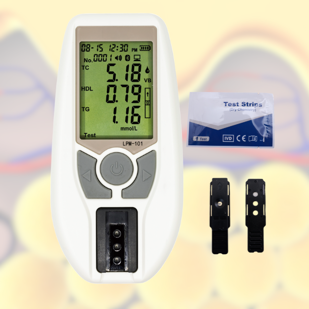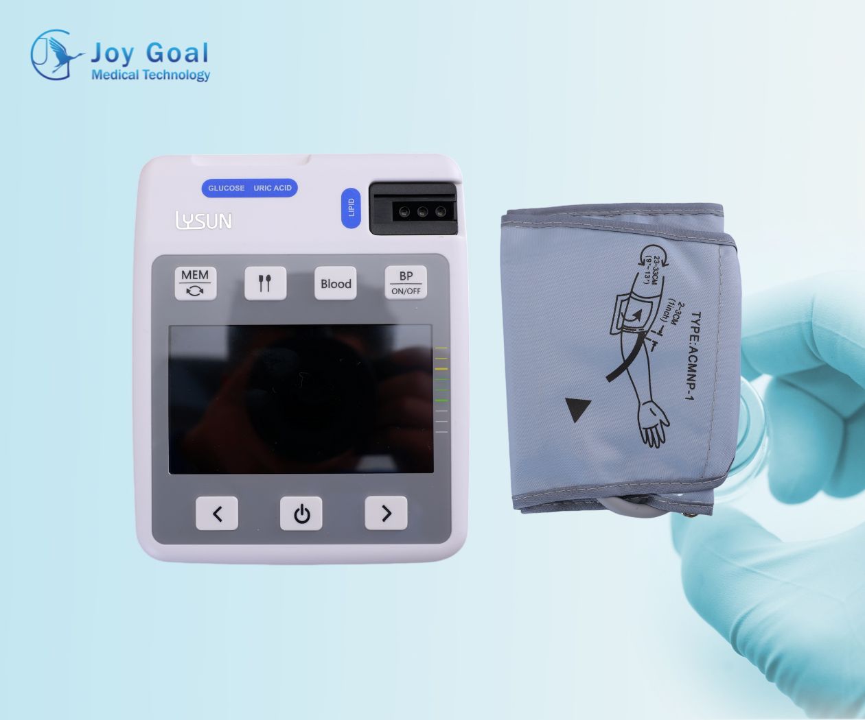
lipid Profile Panel
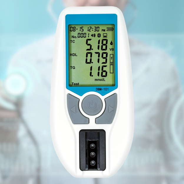
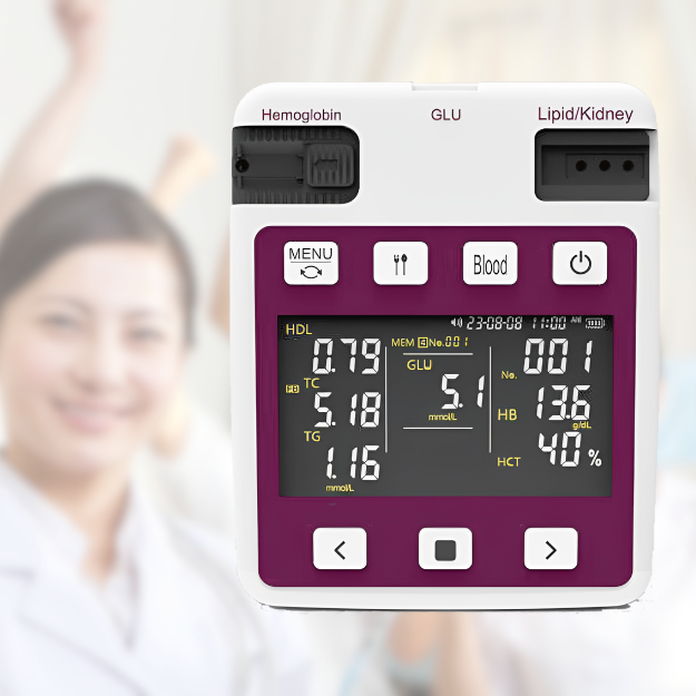
A lipid profile panel is a blood test that measures the amount of lipids (fats) in your blood. These fats include cholesterol and triglycerides. It’s a common test that helps assess your risk of developing cardiovascular disease, such as heart attack or stroke.
Why is it important?
High levels of certain lipids, especially “bad” cholesterol (LDL), can build up in your arteries, forming plaque. This plaque narrows your arteries and restricts blood flow, increasing your risk of heart disease and stroke. Understanding your lipid profile can help you and your doctor make informed decisions about lifestyle changes and, if necessary, medication to manage your cholesterol and triglyceride levels.

Renal Function
Renal function refers to the work performed by your kidneys. These bean-shaped organs play a vital role in maintaining overall health and well-being. They act as your body’s natural filter, constantly working to remove waste products and excess fluids from your blood.
Uric Acid: Elevated levels of uric acid, a condition known as hyperuricemia, can cause gout and lead to kidney stones.
Creatinine:levels in the blood are a marker of how well the kidneys are filtering waste from the blood. Abnormal levels can indicate:
Urea: Elevated levels of urea can indicate kidney impairment because the kidneys are not effectively filtering it from the blood.

Blood hemoglobin
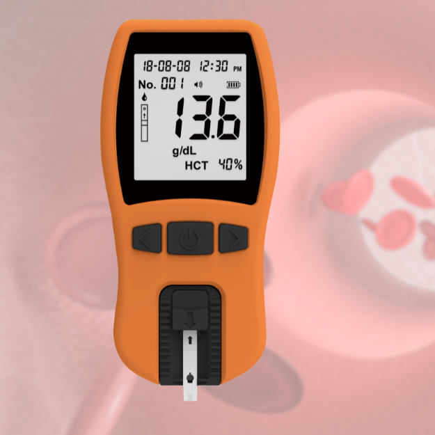
Composition: Hemoglobin is a globular protein composed of four subunits, each containing a heme group. The heme group itself is made up of iron that binds to oxygen atoms. Hemoglobin subunits are known as alpha chains and beta chains (in adults).
Structural Features: The heme group is bound to the protein backbone in a pocket that can bind to one oxygen molecule. In humans, the normal hemoglobin is called hemoglobin A (HbA), and it consists of two alpha and two beta subunits. Other forms of hemoglobin can be present during fetal development (such as fetal hemoglobin, HbF) and in specific genetic conditions.
Function: Each molecule of hemoglobin can carry up to four oxygen atoms. The iron within the heme group is what binds to oxygen. The protein’s structure allows for efficient oxygen release in areas of high carbon dioxide concentration (like muscles during exercise) and low pH (like when carbon dioxide is converted to bicarbonate).

diabetes
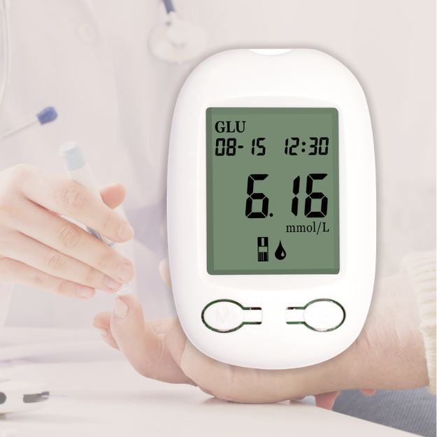
Diabetes, commonly referred to as diabetes mellitus, is a chronic metabolic condition characterized by high blood sugar (glucose) levels resulting from defects in insulin secretion, insulin action, or both. Insulin is a hormone produced by the pancreas that regulates the amount of glucose in the blood. There are several types of diabetes, but the two most common are Type 1 Diabetes and Type 2 Diabetes.
Type 1 Diabetes:
Results from the autoimmune destruction of the beta cells in the pancreas that produce insulin.
Type 2 Diabetes:
Characterized by insulin resistance, where the body’s cells do not effectively respond to insulin.

uric acid
Uric acid is a compound that forms when the body breaks down purines, substances that are found naturally in the body and in certain foods. The process of purine metabolism leads to the production of uric acid, which is then eliminated from the body primarily through the kidneys and to a lesser extent through the digestive tract.
Uric Acid and Health
The levels of uric acid in the blood can have important implications for health. Too much or too little uric acid can lead to various health issues:
Hyperuricemia: This refers to high levels of uric acid in the blood. It can lead to the formation of uric acid crystals that deposit in joints and surrounding tissues, causing gout, a painful form of arthritis. Hyperuricemia can also increase the risk of kidney stones and has been associated with cardiovascular disease.
Hyperuricosuria: This condition involves high levels of uric acid in the urine and is often related to hyperuricemia. It can lead to the formation of uric acid kidney stones.
Hypouricemia: This is less common and refers to abnormally low levels of uric acid in the blood. It is associated with certain disorders such as Lesch-Nyhan syndrome, a rare genetic disease that leads to overproduction of uric acid excretion.

Blood Pressure
Blood pressure is a fundamental vital sign that reflects the force exerted by circulating blood against the walls of blood vessels. It’s a critical measure of cardiovascular health and a key indicator in the diagnosis and monitoring of several health conditions, including hypertension (high blood pressure) and hypotension (low blood pressure).
A healthy blood pressure range for an adult is generally considered to be less than 120/80 mmHg, although individual “normal” ranges can vary.
- Elevated Blood Pressure (120-129/80 mmHg) indicates higher risk of hypertension.
- Hypertension Stage 1 (130-139/80-89 mmHg).
- Hypertension Stage 2 (≥140/≥90 mmHg).
- Hypertensive Crisis (≥180/≥120 mmHg) necessitates immediate medical attention.
Regular blood pressure monitoring at a healthcare facility and sometimes at home is important for detecting and managing hypertension, thus preventing its often serious associated health risks.

SpO2
SpO2, or Pulse Oximetry, is a non-invasive method used to measure the oxygen saturation of a person’s blood, or more specifically, the percentage of oxygen-bound hemoglobin relative to total hemoglobin in the blood. This vital sign is crucial for understanding the efficiency of your lungs in oxygenating the blood and is a critical piece of data in assessing a patient’s respiratory status.
The pulse oximeter, a device used to measure SpO2, shines two wavelengths of light, most commonly red and infrared, through your finger or other body part and measures the absorption of these wavelengths as the light passes through the blood. The oxygenated and deoxygenated hemoglobin in the blood absorb these wavelengths differently, allowing the device to calculate the oxygen saturation percentage.

eCG
An Electrocardiogram, often abbreviated as ECG or EKG (from the German “Elektrokardiogramm”), is a crucial diagnostic tool used in clinical medicine to measure and record the electrical activity of the heart. It’s fundamental in diagnosing heart conditions and health management.
Components of an ECG
P wave: Corresponds to the depolarization of the atria (the heart’s upper chambers).
QRS complex: Represents the depolarization of the ventricles (the heart’s main pumping chambers).
T wave: Shows the repolarization of the ventricles after the contraction.
The ECG is an indispensable tool in cardiology and general medicine, offering a window into the heart’s electrical and functional health. It is essential for the accurate diagnosis and management of various cardiovascular conditions. Understanding ECG patterns can help health professionals tailor treatments and provide personalized care to patients, enhancing medical outcomes.

